 HIPAA Compliant
 ISO 27001 Certified

Breast Biopsy Procedure, types, and what to expect
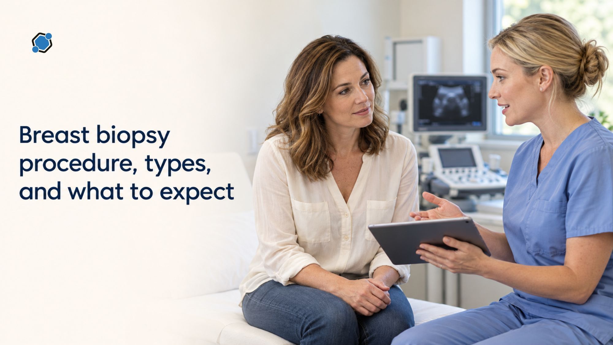
A breast biopsy is a diagnostic step, not a diagnosis. When a mammogram, ultrasound, MRI, or physical exam finds something that needs a closer look, a biopsy removes a small sample of tissue or fluid so a pathologist can examine it under a microscope. According to the Cleveland Clinic, roughly four out of five breast biopsies in women come back benign, meaning no cancer is found. For the remaining cases, the biopsy also provides the tissue needed to guide every treatment decision that follows.
Modern breast biopsies are usually quick, done under local anesthesia, and performed with image guidance in an outpatient clinic. Most take under an hour, and serious complications are rare. The sections below cover why a biopsy is ordered, the main types used today, what happens before, during, and after the procedure, and what the results mean.
Why doctors order a breast biopsy
A clinician recommends a biopsy when imaging or an exam shows something that cannot be fully explained by pictures alone. Common reasons include a suspicious mass or area of calcifications on a mammogram, a lump felt during an exam, a solid area on ultrasound, an abnormal finding on breast MRI, spontaneous or bloody nipple discharge, or persistent skin changes such as dimpling, redness, or crusting.
Imaging shows where an abnormality is and how suspicious it looks, but it cannot always distinguish benign changes from cancer. Only a tissue sample can do that. For a wider look at the warning signs that often lead to further workup, this overview of breast cancer signs beyond a lump covers the symptoms that may prompt imaging and, sometimes, a biopsy.
The main types of breast biopsy
Breast biopsies fall into two broad categories: needle biopsies, which are the standard first-line approach, and surgical biopsies, which are reserved for specific situations. The right method depends on where the lesion is, how it looks on imaging, and the clinician’s judgment.
Fine needle aspiration (FNA)
FNA uses a thin, hollow needle attached to a syringe to withdraw fluid or cells from a cyst, lump, or lymph node. It is the fastest biopsy type, often taking 10 to 15 minutes under local anesthesia. FNA works well for cysts and lymph nodes but collects loose cells rather than tissue structure, so results can sometimes be inconclusive and lead to a follow-up core biopsy.
Core needle biopsy, usually image-guided
Core needle biopsy is the standard way to diagnose most suspicious breast findings today. A larger, hollow spring-loaded needle removes several thin cylinders of tissue through a small skin nick under local anesthesia, in about 20 to 60 minutes. Core samples preserve tissue structure, which lets pathologists distinguish benign changes, non-invasive cancer, and invasive cancer, and run the extra tests that guide treatment.
Most core biopsies are guided by imaging so the needle reaches the exact area of concern. Ultrasound-guided biopsy is often chosen for masses visible on ultrasound and uses no radiation. Stereotactic biopsy uses mammography images from two angles to target calcifications or lesions seen only on a mammogram. MRI-guided biopsy is reserved for findings visible only on breast MRI. Stereotactic and MRI-guided biopsies are often performed with a vacuum-assisted device, which uses gentle suction to collect several contiguous samples through one small opening.
Surgical biopsy
A surgical biopsy is performed in an operating room by a breast surgeon. An excisional biopsy removes the entire abnormal area, often with a small rim of surrounding tissue, while an incisional biopsy removes only part of a larger mass. It may use local anesthesia with sedation or general anesthesia. Surgical biopsy is no longer the first choice for most new findings because needle biopsies are nearly as accurate and far less invasive. It is used when a needle biopsy result does not match the imaging, when certain high-risk lesions are found, or when a needle approach is not technically possible.
Skin punch biopsy
A punch biopsy uses a small circular blade to remove a tiny plug of skin under local anesthesia. It is used for persistent skin changes on the breast, particularly when inflammatory breast cancer or a condition such as Paget disease of the nipple is suspected. In those cases a punch biopsy is usually paired with a core needle biopsy of any underlying tissue.
What happens before, during, and after the biopsy
Preparation for a breast biopsy is usually straightforward. The care team reviews a complete list of medications, including blood thinners, aspirin, NSAIDs, and supplements such as fish oil or vitamin E. Some patients may be asked to pause certain medications for a few days, but no prescribed medication should ever be stopped without the prescribing clinician’s approval. A two-piece outfit is practical, and deodorant, powder, and lotion should be avoided before mammography-based procedures. Eating and drinking are usually permitted before a needle biopsy, but fasting applies when sedation or general anesthesia is planned.
Positioning during the procedure depends on the type. For ultrasound-guided biopsies, the patient usually lies on her back. For stereotactic biopsies, she often lies face-down on a table with the breast through an opening, or sits upright in a dedicated chair. For MRI-guided biopsies, she lies face-down in the scanner. Local anesthetic feels like a brief pinch followed by a sting, and the area numbs within seconds. A small nick is made in the skin, and the biopsy needle is placed with imaging guidance. Patients commonly describe pressure, a clicking or buzzing sound from the sampling instrument, and a sensation of pulling. Sharp pain is uncommon and usually indicates that more anesthetic is needed.
About the marker clip
After the samples are collected, a tiny marker, often titanium or stainless steel and about the size of a sesame seed, is usually placed at the biopsy site through the same needle. This marker, sometimes called a clip, lets future imaging and surgical teams locate the exact spot. According to the American Cancer Society, the marker cannot be felt or seen from the outside, is safe during MRI scans, and will not set off airport metal detectors. If the biopsy shows cancer and surgery is needed, the clip helps the surgeon find the area and is removed with the surrounding tissue. If the result is benign, the clip stays in place safely and permanently.
Recovery and aftercare
After a needle biopsy, firm pressure is applied for several minutes, and the skin opening is closed with adhesive strips or a small dressing. No stitches are typically needed. Patients can usually drive themselves home and return to light activity the same day, though strenuous activity and heavy lifting should generally be avoided for at least 24 to 48 hours. Mild bruising, tenderness, and a small firm area are expected and usually resolve within one to two weeks. The care team should be contacted for expanding swelling, significant bleeding or drainage, warmth or redness, fever, or, after biopsies near the chest wall, sudden chest pain or shortness of breath. These complications are uncommon, each occurring in well under one percent of needle biopsies. Recovery from a surgical biopsy takes longer and involves stitches, a small visible scar, and a few days to a week before normal activity resumes.
What biopsy results mean, and what comes next
Pathology results are usually available within a few business days, though some reports take a week or longer, especially when additional tests are added. Some patients receive a phone call from the ordering clinician, while others see the report first through an online patient portal under federal rules that require rapid release of test results. Deciding in advance whether to review the report alone or wait for the clinician’s call can ease the waiting period.
A pathology report falls into one of several categories. A benign result means no cancer was found, and the radiologist confirms the result matches the imaging. An atypical or high-risk result, such as atypical ductal hyperplasia, is not cancer, but these findings carry a higher risk of cancer being present nearby or developing later, and usually prompt a surgical consultation. A diagnosis of carcinoma in situ, such as ductal carcinoma in situ, means abnormal cells are present but confined. A diagnosis of invasive cancer means abnormal cells have grown into surrounding breast tissue. For a plain-language overview of what each stage designation means, this explainer of breast cancer stages from 0 to 4 walks through how clinicians arrive at a stage after diagnosis.
How biopsy tissue shapes treatment
When invasive breast cancer is found, the pathology lab runs additional tests on the biopsy tissue to characterize the tumor. The three standard biomarkers reported are estrogen receptor (ER), progesterone receptor (PR), and human epidermal growth factor receptor 2 (HER2). These results define the main subtypes and determine which treatments are most likely to help. For a closer look at one of these biomarkers, this breakdown of what HER2 status means for treatment explains how HER2 positive, HER2 negative, and the newer HER2 low category are defined and used. Molecular and genomic testing may also be performed on the biopsy sample to help predict how a tumor is likely to behave.
Common myths, and the role of biopsies in research
One of the most persistent fears is that a needle biopsy can cause cancer to spread. Current evidence does not support this concern. A review published in Cancers in 2024, cited by the American Cancer Society, concluded that needle biopsy does not significantly affect the chances of breast cancer recurrence or long-term survival, and standard treatment addresses the needle path through surgery and radiation when needed. Delaying a recommended biopsy, by contrast, carries real risk because it delays diagnosis.
Other common misconceptions include the belief that a biopsy automatically means cancer, when most results are benign; the fear that the procedure will be extremely painful, when most patients describe pressure and a brief sting rather than sharp pain; and the worry that the marker clip will cause problems with scanners or MRIs, when it is specifically designed to be safe for both. Breast Cancer Awareness Month, observed each October, is a useful anchor for addressing these fears alongside the broader picture of screening and treatment, and this overview of what Breast Cancer Awareness Month covers in 2026 puts biopsy and diagnosis in that wider context.
Beyond diagnosing an individual patient, breast biopsies have long been central to clinical research. Decades of biopsy data and tissue samples have helped identify breast cancer subtypes, develop targeted drugs, and refine how imaging is used. For a wider view of the research and the people driving it forward, this reflection on research and trials behind breast cancer progress highlights the role participants and researchers play in advancing care.
For people recently diagnosed, clinical trials may offer access to therapies not yet broadly available, often selected based on the biomarker results from the biopsy itself. DecenTrialz is a United States-based clinical trial recruitment, pre-screening, and participant management platform that combines AI-driven participant matching with registered-nurse-led pre-screening and structured referral workflows. People share basic health information, the AI matches them to trials that may fit their situation, a registered nurse reviews the match, and a referral is sent to the research site team. Final eligibility determination, informed consent, and enrollment are handled by the research site, not by DecenTrialz. For anyone considering a trial, discussing the option with the treating oncologist is always the first step.
Questions worth asking before the biopsy
Being prepared for the biopsy visit makes the experience less unsettling and ensures the care team can address concerns in advance. A short list of questions can help:
- What type of biopsy is recommended, and why that image guidance method?
- Who performs the procedure, and how long should it take?
- What anesthesia will be used, and is a driver needed afterward?
- Should any medications or supplements be paused, and who confirms the plan?
- How many samples will be taken, and will a marker clip be placed?
- When will results be ready, and will they appear in a patient portal first?
- What are the next steps for each result category, and will biomarker testing be done on this sample if cancer is found?
A breast biopsy can feel like a long day on the calendar, but it is usually a short, well-tolerated procedure with answers that chart the clearest path forward, whether that path leads back to routine screening or to the next step in care.
Was this article helpful?

Similar Blogs
Mobile phlebotomy in clinical trials: how home blood draws work
Blood tests are one of the most common procedures in a clinical trial. They tell the study...
 Anish Teepireddy

Men's Breast Cancer Awareness Week 2026: signs, risks, and the research gap
Every person is born with a small amount of breast tissue, and men are no exception. That ...
 Swaroop ESD

Obesity and Sleep Apnea: How Weight Affects Breathing During Sleep
Excess body weight is the strongest modifiable risk factor for obstructive sleep apnea, a ...
ParamrajStay Informed. Stay Connected.
Get updates on verified clinical trials, emerging treatments, and research breakthroughs directly in your inbox. No spam, just science that matters.
