 HIPAA Compliant
 ISO 27001 Certified

CT vs MRI for stroke: how do doctors choose the right brain scan?
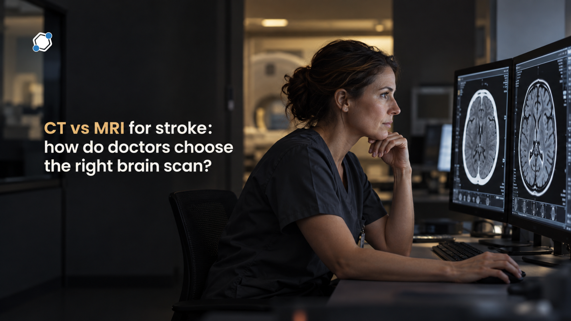
When a stroke is suspected, doctors need a clear picture of the brain before they can safely choose a treatment. The two main options are a CT scan and an MRI, and the question of CT vs MRI for stroke comes down to speed, availability, and how much detail each scan can show. A CT scan is usually the first test in the emergency room because it is fast and reliably shows bleeding. An MRI sees early and small strokes far more clearly, but it takes longer and is not always available right away. Knowing what each scan shows helps patients and families understand the tests ordered during a stroke evaluation.
Why a brain scan comes before stroke treatment
A stroke happens when blood flow to part of the brain is interrupted. Most strokes are ischemic, meaning a clot or narrowed artery blocks blood flow. A smaller share are hemorrhagic, meaning a blood vessel in or around the brain bursts and bleeds. The two types can cause almost identical symptoms, so a physical exam alone cannot tell them apart.
This difference drives every early treatment decision. Clot-dissolving medicines, often called thrombolytics, may restore blood flow in an ischemic stroke, but they could make a bleeding stroke worse. A brain scan is the only reliable way to rule out bleeding before these medicines are considered. The contrast between blocked and burst blood vessels in stroke shapes not only the first scan but the whole treatment path that follows.
Speed matters because brain cells begin to die within minutes when their blood supply stops. Current U.S. stroke guidelines call for brain imaging to be completed within about 25 minutes of a patient's arrival when a stroke is suspected. That goal is one reason the fastest available scan usually comes first.
How a CT scan works and why the ER usually starts with it
A CT (computed tomography) scan uses X-rays and a computer to create cross-sectional images, or slices, of the head. A basic head CT takes only a few minutes, and CT scanners are available around the clock in nearly every U.S. emergency department.
What a CT scan shows well
CT is very good at detecting fresh bleeding in the brain, which stands out clearly on the images. The standard emergency head CT is done without contrast dye, so it can be performed right away without waiting for lab results.
What a CT scan can miss
In the first hours of an ischemic stroke, the affected brain tissue often looks normal on CT. Visible changes can take hours to develop. Small strokes are also hard to see, and so are strokes in the brainstem and the back of the brain, because the thick bone at the base of the skull creates streaks on the images. For these reasons, a normal CT does not rule out a stroke. It mainly shows that there is no major bleeding and no large, established area of damage.
CT angiography and CT perfusion
Many hospitals add two more CT scans that use an iodine-based contrast dye injected into a vein. CT angiography maps the blood vessels of the head and neck and can reveal a large blocked artery. CT perfusion shows how blood is moving through brain tissue. It helps doctors estimate which areas are already permanently damaged and which areas are starved of blood but may still be saved.
These scans help identify patients who may benefit from thrombectomy, a procedure in which a specialist threads a thin tube through an artery to remove the clot. For carefully selected patients, this kind of imaging has extended the time in which clot removal may still be considered, in some cases to many hours after symptoms began.
What an MRI adds to a stroke diagnosis
An MRI (magnetic resonance imaging) scan uses a strong magnet and radio waves, rather than X-rays, to create detailed images of the brain. It does not use ionizing radiation. An MRI takes longer than a CT. A focused emergency stroke MRI may take roughly 10 to 15 minutes, while a full brain MRI often takes 15 to 45 minutes or more.
Seeing a stroke within minutes
A type of MRI image called diffusion-weighted imaging (DWI) detects changes in how water moves inside injured brain cells. A new ischemic stroke can appear on DWI within minutes, long before it becomes visible on CT. In a well-known study comparing the two scans, MRI detected roughly three times as many acute strokes as CT. MRI also detects brain bleeding about as accurately as CT, and it can reveal tiny older bleeds that CT does not show.
Strokes with an unknown start time
Some people wake up with stroke symptoms, or no one saw when the symptoms began. Because treatment decisions depend on timing, this situation once ruled out clot-dissolving medicine for many patients. MRI can help estimate how recent a stroke is by comparing two image types. When a stroke appears on DWI but not yet on a second image type called FLAIR, it likely began within the past few hours. Current guidelines consider this approach reasonable for selected patients whose start time is unknown.
Mini-strokes and small strokes
MRI is the preferred scan for evaluating a transient ischemic attack (TIA), often called a mini-stroke, ideally within 24 hours. The symptoms of a TIA go away, but MRI sometimes shows a small area of injury anyway. That finding signals a higher risk of a future stroke and may change the follow-up plan. Recognizing the warning signs of a mini-stroke and seeking care quickly makes this kind of early imaging possible.
Where MRI has limits
MRI is not always available at night, on weekends, or in smaller and rural hospitals. The scanner is loud and enclosed, which can be difficult for people with claustrophobia, and patients must lie still, which can be hard during a stroke. Metal in the body also requires screening. Many newer pacemakers, defibrillators, and other implants are labeled MR conditional, meaning they can be scanned safely under specific conditions, but some implants are not safe in the scanner. It also generally costs more than CT.
CT vs MRI for stroke: how the two scans compare
Neither scan is better in every situation. Each answers different questions at different points in a stroke evaluation, and current guidelines accept either one as the first scan, as long as it is done quickly and does not delay treatment.
Speed and availability
CT is faster and nearly always available. MRI takes longer and may not be ready immediately, especially outside regular hours. In an emergency where minutes count, this gap often decides which scan comes first.
Accuracy
Both scans detect acute bleeding reliably. MRI is far more sensitive for ischemic strokes in their first hours, for small strokes, and for strokes in the brainstem, all of which CT can miss.
Radiation and safety
A standard head CT delivers about as much radiation as roughly seven months of natural background radiation from the environment. Adding angiography and perfusion scans raises the total. MRI uses no ionizing radiation but requires screening for metal and implants. Both tests sometimes use contrast dye, and the care team weighs allergies and kidney function. Even so, guidelines advise against delaying urgent blood vessel imaging just to wait for kidney test results.
In practice, many people with a suspected stroke have a CT first, sometimes with angiography and perfusion, and then an MRI later during the hospital stay. The MRI can confirm the diagnosis, show the exact size and location of the stroke, and help guide prevention.
How brain imaging shapes stroke research
Brain scans play a central role in stroke clinical trials. Many studies use imaging to decide who can take part, for example by requiring a blocked large artery, a certain amount of brain tissue that may still be saved, or a specific pattern on MRI. Scans are also used to measure results, such as whether the area of damage grows or whether bleeding occurs after treatment.
Because of this, the timing and type of a person's scans can affect whether a study is a good fit. These requirements protect participants and help researchers produce reliable results. Imaging criteria are one of several reasons people can be screened for a study and still not enroll.
DecenTrialz is a clinical trial recruitment and pre-screening platform that helps people find stroke studies that may fit their situation. People share some information about their health, AI-assisted matching surfaces relevant studies, and a registered nurse reviews the information and explains what each study involves. Interested individuals are then referred to the research site team.
The research site team makes all final eligibility decisions, handles informed consent, and manages enrollment. DecenTrialz does not interpret scans, give medical advice, or guarantee access to any study. Stroke survivors and caregivers who want to learn about research options can explore current studies through DecenTrialz.
Common questions about CT and MRI for stroke
Can a CT scan miss a stroke?
Yes. An ischemic stroke in its first hours, a small stroke, or a stroke in the brainstem may not show up on CT. A normal CT mainly rules out major bleeding. Doctors may order CT angiography, CT perfusion, or an MRI when symptoms still point to a stroke.
Why did the ER do a CT instead of an MRI?
The first priority is to rule out bleeding within minutes so treatment decisions can be made quickly. CT is usually the fastest scan available at any hour. An MRI may follow later to add detail.
Does a silent stroke show up on MRI?
Often, yes. Silent strokes are areas of brain injury found on imaging in people who never noticed stroke symptoms. MRI is especially good at revealing them, and they are often discovered when a brain scan is done for another reason. An overview of silent stroke signs and long-term effects explains why these findings matter.
Is an MRI safe for people with a pacemaker or metal implant?
It depends on the device. Many modern devices are MR conditional and can be scanned safely when specific conditions are followed. The care team needs to know the exact type of any implant before the scan.
Can a stroke be seen on MRI weeks or months later?
Usually, yes. Most completed strokes leave a lasting mark that standard MRI images can show long after the event. The signal that identifies a fresh stroke fades over time, however, and very small changes may become harder to see.
Why calling 911 matters more than which scan comes first
The choice between CT and MRI belongs to the emergency care team. What matters most for a person having a stroke is reaching a hospital quickly, because treatment options narrow with every passing minute. Knowing the BE FAST warning signs of stroke helps bystanders act without delay:
- B is for Balance: sudden loss of balance or coordination.
- E is for Eyes: sudden blurred vision or vision loss in one or both eyes.
- F is for Face: one side of the face droops or feels numb.
- A is for Arms: one arm is weak or numb.
- S is for Speech: slurred speech or trouble finding words.
- T is for Time: call 911 right away.
Calling 911, rather than driving to the hospital, allows emergency medical services to begin care and alert the hospital stroke team before arrival. Anyone who notices these signs in themselves or someone else should call 911 immediately, even if the symptoms go away.
Was this article helpful?

Similar Blogs

How do digital endpoints in clinical trials use data from wearable devices?
Digital endpoints in clinical trials are measurements of health that are collected by a de...
 Deeksha Gitta

BRCA1 and BRCA2 mutations: what they mean for breast cancer risk
BRCA1 and BRCA2 mutations are inherited gene changes that raise the risk of breast, ovaria...
Swaroop ESD
Obesity and fatty liver: how does extra weight damage the liver?
Obesity is the strongest known driver of fatty liver disease, a condition in which excess ...
Deeksha GittaStay Informed. Stay Connected.
Get updates on verified clinical trials, emerging treatments, and research breakthroughs directly in your inbox. No spam, just science that matters.
